Cartilage Lesions

The cartilage that coats the knee articulation facilitates low friction and painless mobility between the various articular surfaces. However, its wear can be the cause of knee pathologies.

Chondromalacia patellae (CMP)

Chondromalacia patellae (CMP) is a degenerative joint disease that is frequent in female teenagers. It is characterised by the softening (malacia) and fissuring of the cartilage surface. The most common symptom is pain while running and going up or downstairs. Physiotherapy and hydrotherapy in particular are essential in CMP treatment.
Localised Chondral Lesions

Localised Chondral Lesions

Localised chondral lesions (such as osteochondritis dissecans or osteochondral fractures, etc.) usually affect the subchondral. Bone and arthroscopic interventions are thus required to regularise these injuries (shavings and curettages), to revascularise them (pridie drilling, retrograde drilling, micro fractures) or to replace the injured cartilage with healthy one (grafts) from other parts of the knee (mosaicplasty).

Recently, there have been attempts to use platelet-derived growth factor (PDGF) and chondrocytes culture (MACI) to treat these pathologies, techniques that we have been testing along with the use of stem cells.
Patello-Femoral Syndromes

Patello-Femoral Syndromes

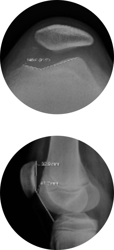
The Patello-Femoral Syndrome's aetiology usually lies in the deficient alignment of the knee's extensor apparatus, which causes the side tilting of the patella when the quadriceps is contracted, leading to hyperpressure in the external side of the patella and eventually to instability and recurrent dislocations. Imaging exams (in this particular case, X-Ray, CAT and MRI Scanning) are required to study the patellofemoral interactions, to measure the patellar height, the trochlear inclination angle, the patellar tilt angle, TT-TG, etc.

The symptoms and the results of the exams will help determine the course of treatment.

In general, for patients with Patello-Femoral Syndromes where the alignment between the tuberosity of the tibia and the trochlear groove is relatively well preserved, physiotherapy to strengthen the medial vastus is indicated. This goal can be achieved with isometric or isotonic exercises performed with the knee in external rotation or even with electro-stimulation, which helps to develop the target muscle group.

If imaging test results show angle alterations exceeding standard values, then a proximal (medial patellofemoral ligament reconstruction) or distal realignment surgery (with medialisation of the tibial tuberosity) is required.